🫁❤️🚴♂️📈 How the Heart, Lungs, and Muscles Work Together During CPET
🩺 Understanding the Three Gears in Exercise Physiology
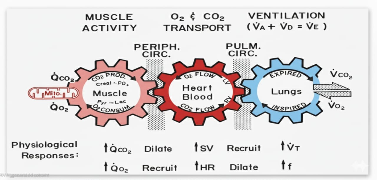
This concept beautifully explains how the muscles, heart, and lungs work together during exercise. Each acts like a gear — when one moves, the others turn with it. If one slows down, the whole system is affected.
During a Cardiopulmonary Exercise Test (CPET), we measure how efficiently these three systems coordinate in real time.
1. Muscle Activity — Where the Demand Begins
It all starts at the muscle. Inside the mitochondria, oxygen (QO₂) is used to generate ATP, while carbon dioxide (QCO₂) is produced as a metabolic by-product.
As workload increases, muscles consume more oxygen and produce more CO₂. This rising metabolic demand signals the cardiovascular system to deliver more oxygenated blood.
Local vasodilation occurs around active muscles — blood vessels widen to increase flow, ensuring oxygen delivery and CO₂ removal keep pace with demand.
2. O₂ and CO₂ Transport — The Heart as the Middle Gear
The heart links the lungs and muscles. Oxygenated blood from the lungs enters the left ventricle and is pumped to the working muscles, while deoxygenated blood returns via the right ventricle for gas exchange.
During exercise, the heart increases:
- Stroke volume (SV)
- Heart rate (HR)
Together, these raise cardiac output (Q), matching the rising metabolic demand.
Early in exercise, stroke volume increases through better ventricular filling and stronger contraction. As intensity rises, heart rate becomes the main driver.
Blood vessels also dilate to accommodate the rising flow, helping maintain balance between oxygen delivery and carbon dioxide clearance.
3. Ventilation — The Final Gear
The lungs complete the circuit. Here, oxygen enters the blood while CO₂ is exhaled.
With rising intensity:
- Tidal volume (VT) increases first
- Then breathing frequency (f) rises
This expands total ventilation (VE), supporting the matching of oxygen uptake (VO₂) and carbon dioxide elimination (VCO₂), even during high metabolic demand.
4. Physiological Recruitments Behind the Gears
At the bottom of the diagram, we see how each system reacts when demand rises:
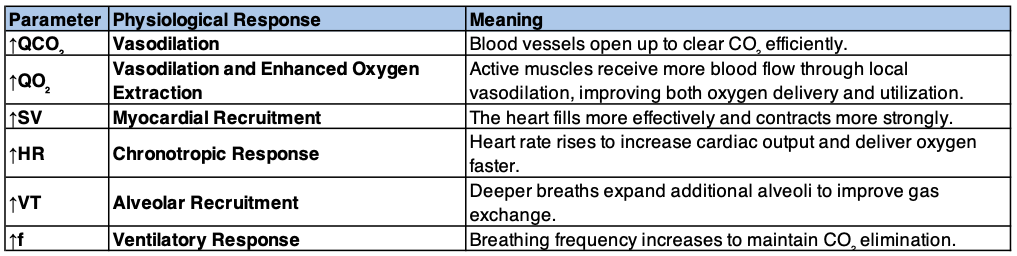
Each of these adjustments ensures that muscles receive what they need at the right time. When one mechanism fails — say, if cardiac output doesn’t rise enough or the lungs can’t clear CO₂ efficiently — the other systems try to compensate. This is often how CPET helps pinpoint the exact level of limitation.
If one mechanism underperforms — such as low cardiac output or impaired ventilation — the others try to compensate. This interplay often reveals the exact point of limitation during CPET.
5. Why CPET Captures It All
CPET measures every component of this integrated response:
- Oxygen uptake (VO₂)
- Carbon dioxide output (VCO₂)
- Ventilation (VE)
By analysing these variables together, CPET identifies which “gear” is underperforming:
- Cardiac limitation → inadequate oxygen delivery
- Pulmonary limitation → reduced ventilatory efficiency
- Muscular limitation → early fatigue due to metabolic inefficiency
Summary
Exercise physiology is teamwork between the muscles, heart, and lungs. CPET lets us observe how this teamwork behaves under stress — not in isolation, but as a synchronised system.
When one gear slows down, CPET helps pinpoint exactly where and why.
📖 References
- Wasserman, K. et al. (2020). Principles of Exercise Testing and Interpretation. Wolters Kluwer.
- Andonian, B.J. et al. (2022). Making CPET Interpretable for Clinicians. Frontiers in Physiology.
- Chambers, D.J., Wisely, N.A. (2019). CPET — A Beginner’s Guide to the Nine-Panel Plot. BJA Education.
- Stringer, W.W. (2019). Physiological Determinants of Exercise Capacity. Clinics in Chest Medicine.
- Palange, P. et al. (2007). Clinical Exercise Testing and Lung Diseases. European Respiratory Journal.